lab manual for anatomy and physiology
Discover a comprehensive anatomy and physiology lab manual with interactive guides, step-by-step exercises, and visual aids to enhance your learning experience.
The Laboratory Manual for Anatomy and Physiology is a comprehensive resource designed for a two-semester course. It features dynamic activities and experiments to help students visualize anatomical structures and understand physiological concepts. This manual provides a hands-on approach to learning, making complex topics engaging and accessible for all learners.
1.1 Importance of Laboratory Study in Anatomy and Physiology
Laboratory study is essential for understanding anatomy and physiology, as it provides hands-on experience with anatomical structures and physiological processes. Labs allow students to visualize and interact with specimens, enhancing their comprehension of complex topics. Through experiments and dissections, students develop critical thinking and observational skills. Practical exposure to tissues, organs, and systems bridges the gap between theory and real-world applications, preparing learners for careers in healthcare and scientific research. Lab activities also foster collaboration and problem-solving, making them a cornerstone of anatomy and physiology education.
1.2 Key Terminology and Concepts
Key terminology in anatomy and physiology includes terms like anatomy (study of structure) and physiology (study of function). Understanding concepts like homeostasis, cells, tissues, organs, and systems is foundational. The hierarchical organization of the body, from cells to systems, is crucial. Terms like feedback mechanisms and regulation explain how the body maintains balance. Mastery of this terminology and concepts is essential for analyzing laboratory experiments and understanding the human body’s intricate functions and interrelationships.
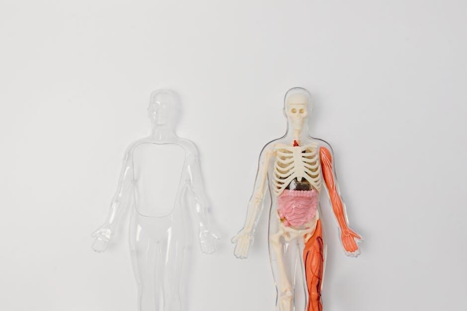
1.3 Overview of Body Systems
The human body is composed of 11 major interconnected systems, each with unique functions essential for overall health; These include the integumentary, skeletal, muscular, nervous, circulatory, respiratory, digestive, urinary, reproductive, endocrine, and immune systems. Each system interacts to maintain homeostasis and ensure proper bodily functions. The lab manual provides detailed exercises to explore these systems, from identifying bones and muscles to studying nerve tissues and organ structures, offering a hands-on approach to understanding their roles and interdependencies.
Laboratory Safety and Equipment
Laboratory safety is crucial for protecting students and ensuring a successful learning environment. Essential equipment includes microscopes, dissection tools, and protective gear like gloves and goggles. Proper handling of specimens, chemicals, and instruments is emphasized to prevent accidents; Students are encouraged to familiarize themselves with lab tools and follow established safety protocols to maintain a clean and organized workspace. Understanding lab equipment and adhering to safety guidelines are fundamental skills for conducting effective anatomy and physiology experiments.
2.1 Safety Protocols in the Anatomy and Physiology Lab
Safety protocols are essential in the anatomy and physiology lab to protect students and ensure a secure learning environment. Proper use of personal protective equipment (PPE), such as gloves and goggles, is mandatory. Students must handle biological specimens, chemicals, and sharp instruments with care to avoid accidents. Clearing workstations and disposing of waste correctly are critical. Emergency procedures, like knowing the location of fire extinguishers and first aid kits, are emphasized. Adhering to these guidelines ensures a safe and effective laboratory experience for all participants.
2.2 Essential Laboratory Equipment and Tools
Laboratory equipment and tools are vital for conducting anatomy and physiology experiments. Common tools include microscopes for examining tissues, dissection instruments like scalpels and forceps, and measurement devices such as calipers and thermometers. Specimen storage solutions, like Formalin and labeled containers, are also essential. Safety gear, including gloves and goggles, protects students during procedures. Additionally, lab manuals and digital tools, such as tablets for accessing e-texts, enhance the learning experience. These resources ensure accurate and efficient completion of lab activities while maintaining a safe environment.
Histology and Microscopy
Histology and microscopy involve the study of tissues and cells under a microscope. This section of the lab manual includes exercises on preparing and examining tissue samples, emphasizing microscopic techniques to understand cellular structures and their functions in various body systems.
Histology is the study of tissue structure, focusing on how cells organize into functional units. This section introduces students to the fundamentals of histology, including tissue classification and microscopic techniques. Through hands-on activities, learners develop practical skills in preparing and staining tissue samples. The lab manual emphasizes the importance of histology in understanding human health and disease, providing a foundation for advanced studies in anatomy and physiology. Key concepts include the use of microscopes, tissue preparation methods, and the identification of cellular structures.
3.2 Preparing and Examining Tissue Samples
Preparing and examining tissue samples is a critical skill in histology. Students learn to fix, stain, and mount tissues for microscopic analysis. The lab manual guides learners through safety protocols, such as handling chemicals and using microscopes. Activities include identifying cellular structures and understanding tissue organization. Practical exercises cover both plant and animal tissues, emphasizing the link between tissue structure and body function. This hands-on approach reinforces theoretical concepts and develops observational skills essential for anatomy and physiology studies.
Integumentary System
The integumentary system, including the skin, hair, nails, and associated glands, is explored through detailed lab exercises. Students examine its structure, function, and protective roles in the body.
4.1 Structure and Function of the Skin
The skin, the body’s largest organ, consists of three primary layers: the epidermis, dermis, and hypodermis. The epidermis, the outermost layer, provides protection against external factors. The dermis contains blood vessels, nerve endings, and glands, while the hypodermis anchors the skin to underlying tissues. The skin’s functions include protection, regulation of body temperature, sensation, and secretion through sweat glands. Lab exercises explore these structures and functions, offering hands-on insights into the skin’s vital roles in maintaining homeostasis and overall health.
4.2 Lab Exercises on the Integumentary System
Lab exercises on the integumentary system provide hands-on exploration of skin structure and function. Activities include examining histological slides of skin layers, identifying touch receptors, and analyzing the effects of environmental factors. Students may also conduct experiments simulating wound healing and burns, observing the body’s repair mechanisms. Practical exercises reinforce theoretical knowledge, enabling a deeper understanding of the skin’s protective, regulatory, and sensory roles. These labs prepare students for real-world applications in healthcare and research, emphasizing the integumentary system’s critical functions in maintaining overall health.
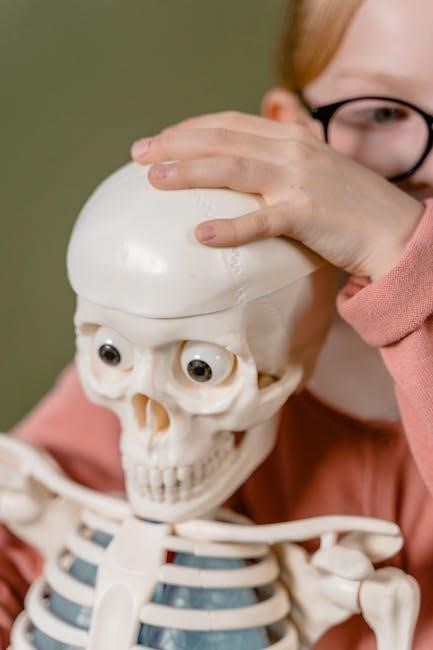
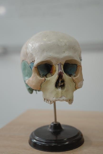
Skeletal System
The skeletal system lab explores the structure and function of bones and joints. Exercises include identifying bone types, examining joint mechanics, and understanding skeletal movements through hands-on activities.
5.1 Bones and Joints
The skeletal system consists of 206 bones, classified into long, short, flat, irregular, and sesamoid types. Bones provide structural support, protect internal organs, and facilitate movement. Joints, or articulations, are points where bones meet, allowing for mobility. The lab manual includes exercises to identify bone structures, such as the femur and tibia, and to examine joint types, like synovial and cartilaginous joints. Students learn to distinguish between diarthrodial joints, which allow wide ranges of motion, and fibrous joints, which are immovable. These activities enhance understanding of skeletal anatomy and its role in movement and stability.
5.2 Lab Activities on the Skeletal System
Lab activities focus on exploring the skeletal system through hands-on exercises. Students identify and analyze bones, such as the femur and tibia, noting their structures and functions. Joint dissection and examination highlight the differences between synovial, cartilaginous, and fibrous joints. Activities include mapping bone markings and simulating joint movements to understand mobility and stability. Interactive tools, like 3D models and digital simulations, enhance learning. These exercises provide a practical understanding of skeletal anatomy, preparing students for advanced studies in musculoskeletal physiology and related healthcare fields.
Nervous System
The nervous system is explored through detailed lab activities, focusing on neural structures and functions. Hands-on exercises include examining brain and spinal cord specimens, identifying neurons, and tracing nerve pathways. These activities provide a foundational understanding of the nervous system’s role in controlling body functions and responding to stimuli, essential for advanced studies in neuroscience and physiology.
Neuroanatomy focuses on the structure and organization of the nervous system, including the brain, spinal cord, and peripheral nerves. Lab exercises introduce students to neural tissues, emphasizing the identification of neurons, glial cells, and synaptic connections. Hands-on activities involve examining histological slides of brain regions, such as the cerebrum and cerebellum, to understand their functions. Students also explore reflex pathways and sensory-motor relationships, gaining insights into how the nervous system controls body functions and responds to stimuli. These foundational studies are crucial for understanding neurological processes and disorders.
6.2 Lab Experiments on Neural Structures
Lab experiments on neural structures provide hands-on opportunities to explore the nervous system’s components. Students examine histological slides of brain and spinal cord tissues, identifying key neural features under microscopes. Activities include dissecting nerve specimens to observe structural organization and conducting reflex testing to demonstrate neural pathways. Digital tools and simulations complement these exercises, offering 3D visualizations of neural connections. These experiments enhance understanding of synaptic transmission, sensory-motor responses, and the nervous system’s integrative functions, preparing students for advanced studies in neuroscience and related fields.
Muscular System
The muscular system is explored through detailed lab activities, focusing on the structure and function of skeletal, smooth, and cardiac muscles. Experiments emphasize muscle physiology.
7.1 Types of Muscles and Their Functions
The lab manual explores the three types of muscles: skeletal, smooth, and cardiac. Skeletal muscles enable voluntary movement and support posture, while smooth muscles handle involuntary actions like digestion. Cardiac muscles pump blood efficiently. Labs include identifying muscle tissues under microscopes and studying their physiological roles. Hands-on activities, such as palpation and dissection, reinforce understanding of muscle structure and function, preparing students for clinical applications.
7.2 Lab Activities on Muscle Physiology
Lab activities focus on understanding muscle function through hands-on experiments. Students explore muscle contraction mechanisms using nerve stimulation and histological slides. Palpation exercises help identify major muscle groups, while physiological experiments measure contraction force. Digital tools, like data acquisition systems, record muscle activity, enhancing understanding of neuromuscular interactions. These activities reinforce muscle physiology concepts, providing practical insights into how muscles contribute to movement and overall bodily functions. Interactive labs ensure a comprehensive learning experience, bridging theory with real-world application.

Senses and Special Senses
This section explores the anatomy and physiology of sensory systems, focusing on vision, hearing, and other special senses. Lab activities include examining eye and ear structures, conducting sensory perception experiments, and understanding how these systems function. Interactive exercises and digital tools enhance learning, helping students connect sensory structures with their physiological roles in perception and response.
8.1 Anatomy of the Eye and Ear
The eye and ear are complex sensory organs with specialized structures for detecting light and sound. The eye consists of the cornea, lens, retina, and optic nerve, working together to focus images and transmit visual signals. The ear includes the outer ear, eardrum, ossicles, and cochlea, converting sound vibrations into neural signals. Lab exercises involve dissecting these organs to identify key anatomical features and understanding their functional roles in vision and hearing. Interactive diagrams and microscopy enhance comprehension of these intricate systems.
8.2 Lab Exercises on Sensory Physiology
Lab exercises on sensory physiology provide hands-on exploration of how the body detects and interprets stimuli. Activities include testing visual acuity, analyzing color perception, and examining hearing thresholds. Students use tuning forks to demonstrate sound wave transmission and conduct nerve response experiments. Practical exercises involve dissecting sensory organs, such as the eye and ear, to identify structures like the retina and cochlea. These labs enhance understanding of how sensory systems convert external stimuli into neural signals, enabling perception and response. Interactive simulations and data analysis further reinforce key physiological concepts.
Digestive System
The digestive system includes the mouth, esophagus, stomach, and intestines, functioning to break down food into nutrients for absorption and energy. Labs explore mechanical and chemical digestion processes.
9.1 Structure and Function of the Digestive Tract
The digestive tract, also known as the alimentary canal, is a tube-like structure extending from the mouth to the anus. It includes the esophagus, stomach, small intestine, and large intestine. The walls of the digestive tract are composed of four layers: mucosa, submucosa, muscularis, and serosa. The mucosa lining secretes enzymes and absorbs nutrients, while the muscularis facilitates peristalsis, moving food through the tract. The digestive tract’s primary functions include ingestion, mechanical and chemical digestion, absorption of nutrients, and elimination of waste. Lab exercises often involve studying the histology of these layers and their roles in digestion.
9.2 Lab Experiments on Digestive Processes
Laboratory experiments on the digestive system focus on understanding its functional mechanisms. Activities include analyzing the role of gastric juice in protein digestion, observing the effects of pH on enzyme activity, and simulating digestive processes in a controlled environment. Students often conduct experiments using histological slides of the digestive tract to identify structural adaptations. Additionally, enzyme assays and pH testing are performed to demonstrate how the digestive system breaks down nutrients. These hands-on exercises provide practical insights into the physiology of digestion, enhancing comprehension of the digestive tract’s role in maintaining bodily functions.
Respiratory System
The respiratory system is explored through lab exercises that examine lung structure, gas exchange, and breathing mechanics. Students analyze tracheal and bronchial tissues to understand airflow pathways.
10.1 Anatomy of the Respiratory System
The respiratory system consists of the upper and lower airways, including the nasal cavity, pharynx, larynx, trachea, bronchi, and lungs. The nasal cavity filters, warms, and humidifies air, while the pharynx serves as a shared pathway for air and food. The larynx contains vocal cords, enabling speech and preventing foreign particles from entering the trachea. The trachea divides into bronchi, which branch into bronchioles leading to alveoli, where gas exchange occurs. Lab exercises involve dissecting lung tissue and observing the microscopic structure of alveoli to understand their role in respiration. The diaphragm and intercostal muscles facilitate breathing movements, essential for oxygen intake and carbon dioxide expulsion.
10.2 Lab Activities on Breathing Mechanics
Lab activities on breathing mechanics focus on understanding the physiological processes of respiration. Students use spirometry to measure lung capacities, such as tidal volume and vital capacity, and analyze breathing patterns. Exercises include observing chest and abdominal movements to assess diaphragm function. Simulation models demonstrate the role of intercostal muscles in inhalation and exhalation. Practical experiments, such as measuring respiratory rates and volumes, help quantify respiratory efficiency. These hands-on activities bridge theoretical knowledge with real-world applications, enhancing comprehension of how the respiratory system maintains homeostasis through precise mechanical functions.
Cardiovascular System
The cardiovascular system is explored through detailed lab activities, focusing on the structure-function relationship of the heart and blood vessels. Practical experiments enhance understanding of blood circulation mechanics.
11.1 Heart and Blood Vessel Anatomy
The cardiovascular system is explored through detailed lab activities, focusing on the structure-function relationship of the heart and blood vessels. Practical experiments enhance understanding of blood circulation mechanics. The lab manual includes dissection guides, detailed illustrations, and activities to identify key structures such as heart chambers, valves, and blood vessel types. Students learn to distinguish between arteries, veins, and capillaries, and explore how these components work together to maintain blood flow and overall health. Interactive exercises and visual aids, like those from Marieb/Hoehns, provide a comprehensive learning experience.
11.2 Lab Experiments on Blood Circulation
Laboratory exercises focus on understanding blood circulation through hands-on activities. Students measure blood pressure using sphygmomanometers and analyze pulse rates to observe cardiovascular responses. Simulations and digital tools demonstrate blood flow dynamics and oxygen transport mechanisms. Exercises include studying blood vessel structure and function, as well as the effects of factors like exercise and stress on circulation. These experiments provide practical insights into how the cardiovascular system maintains homeostasis and adapts to physiological changes, reinforcing theoretical concepts with real-world applications.

Additional Resources and Tools
Digital lab manuals and e-Labs provide interactive learning experiences, while online platforms offer supplementary materials, videos, and quizzes to enhance anatomy and physiology studies. These tools promote engagement and understanding.
12.1 Digital Lab Manuals and e-Labs
Digital lab manuals and e-Labs offer interactive and flexible learning experiences for anatomy and physiology students. These resources provide access to virtual dissections, 3D models, and multimedia content, enabling students to explore complex structures and processes in detail. Many digital manuals are optimized for mobile devices, allowing learners to study anytime, anywhere. They often include self-assessment quizzes, interactive diagrams, and videos to reinforce concepts. Additionally, some platforms offer customizable content, enabling instructors to tailor labs to their course needs. These tools enhance traditional learning and cater to diverse learning styles, making anatomy and physiology more engaging and accessible.
12.2 Online Anatomy and Physiology Learning Platforms
Online learning platforms for anatomy and physiology provide interactive tools and resources to enhance student engagement. Platforms like Pearson’s eText and others offer 3D models, virtual simulations, and interactive quizzes to aid in understanding complex concepts. Many platforms integrate with lab manuals, allowing students to access materials anytime. These resources often include self-assessment tools and tracking features to monitor progress. They cater to diverse learning styles and complement traditional lab experiences, making anatomy and physiology study more flexible and effective for modern learners.
The Lab Manual for Anatomy and Physiology is a vital resource for understanding human anatomy and physiology. It offers a comprehensive and practical approach to learning, emphasizing continuous exploration and mastery of physiological concepts.
13.1 Summary of Key Concepts
The Lab Manual for Anatomy and Physiology effectively summarizes the study of the human body’s structure and function. It covers anatomical terminology, histology, and the integumentary, skeletal, nervous, and muscular systems. The manual emphasizes hands-on learning through experiments and dissections, reinforcing understanding of complex physiological processes. By integrating visual aids and practical exercises, it bridges theory with real-world applications, making it an essential tool for students and healthcare professionals alike. This comprehensive approach ensures a solid foundation in anatomy and physiology, fostering a deeper appreciation of human biology.
13.2 Importance of Continuous Learning in Anatomy and Physiology
Continuous learning in anatomy and physiology is crucial due to the evolving nature of scientific discoveries and medical advancements. As new technologies and research emerge, staying updated ensures a deeper understanding of human biology. Laboratory manuals and digital resources provide hands-on experiences, fostering a lifelong commitment to learning. By engaging with updated materials and practical exercises, students and professionals can refine their skills and adapt to emerging trends. This ongoing process enhances both academic and professional growth, making it essential for anyone in healthcare or biological sciences.